These diagrams are to be used for the purpose of education. Within a classroom setting, teachers can either use the simplified depictions of the reproductive system as a viewable diagram to teach anatomy, or as an interactive learning tool. Each diagram includes a simplified drawing of the general organs, one with labeled boxes, and one with a fill-in-the-blank capability. The labeled diagram can be used as an answer key, or as a handout that students can use as a visual aid. The simplistic style of the drawings is meant to allow for easier understanding of these systems, and they can even be colored in as part of an assignment. These diagrams include several key anatomy features, but can be altered to include either more or less detail/information, depending on the teacher’s curriculum.
These diagrams include:
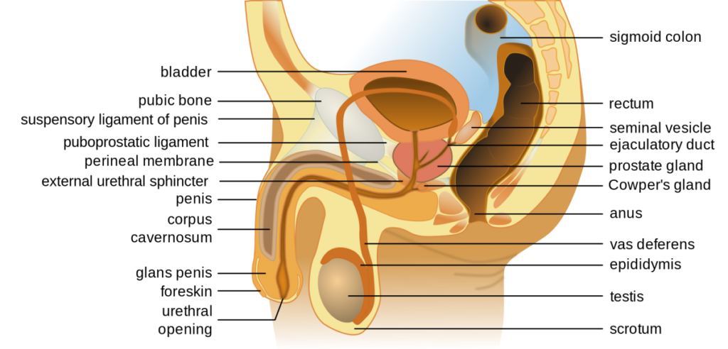
- The internal male reproductive system as viewed from the side(cross section)
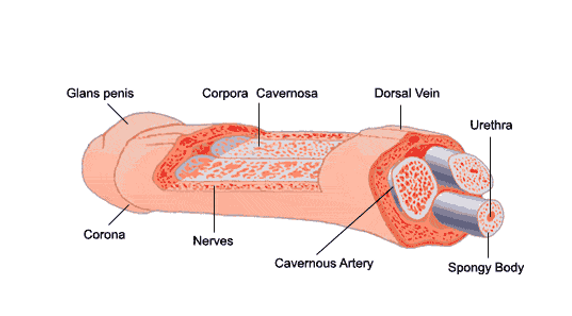
- The external male reproductive system, including a penis, with a ‘cut out’ to see the inside, and close-ups of a cross section of a penis, and a cross section of a testicle
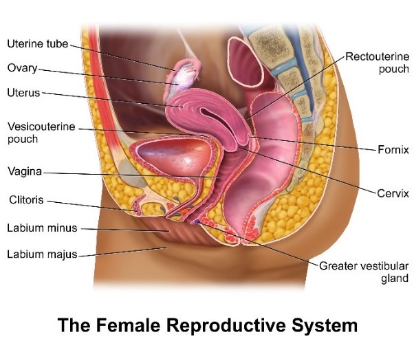
- The internal female reproductive system as viewed from the front
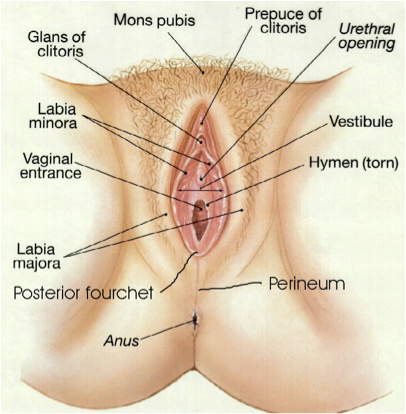
- The external female reproductive system as viewed from the bottom, including the vulva
Table of Contents
The Vulva:
Male Reproductive System (Side View):
Penis/Cross section:
Female Inner Reproductive System: