
Disclaimer: We acknowledge that not all pregnant females will remain the mother of the child and that not all mothers may have biologically given birth to their child. In this article, we use the term “mother” for the sake of simplicity and consistency.
Table of Contents
Overview
Pregnancy lasts about forty weeks when counted from the first day of your last normal period. These weeks are separated into three trimesters. Trimester one spans from weeks 1 to 12. Trimester two is from weeks 13 to 26. Trimester three is from weeks 27 to 40. This article will explain the changes that the mother and her baby experience during these three stages!
The mother and fetus will experience changes each week of the pregnancy. The changes mentioned in this article are all normal and expected for fetuses and their mothers. It is important to remember that healthy babies come in a variety of sizes and can sometimes reach milestones at different times. The baby growth in inches and ounces mentioned in this article refers to average sizes. If a female is pregnant and spotting, having abdominal cramps, or experiencing intensified forms of the symptoms listed below, she should seek medical help immediately. A mother should also be sure to get regular examinations during her entire pregnancy.
1st Trimester (Weeks 1 to 12)
During the first twelve weeks of pregnancy, a female’s body begins to change as the fetus starts to develop. The mother will generally have her first ultrasound or sonogram around the sixth or eighth week of pregnancy.
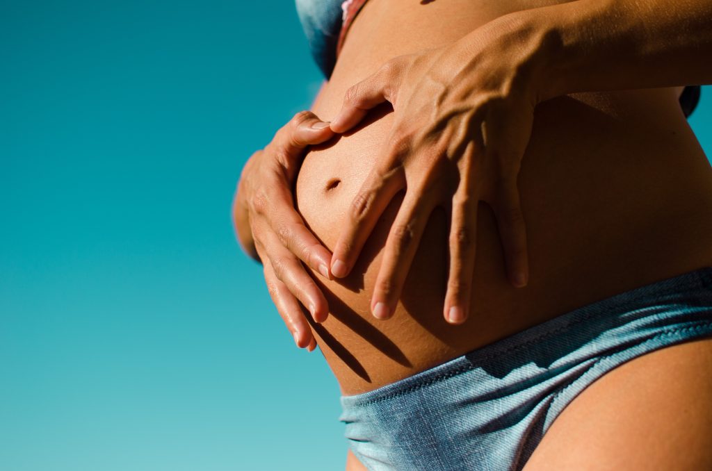
1st Trimester: Pregnant Female
During the 1st trimester, a female may not even know she is pregnant. The first sign of pregnancy that she may encounter is her missed period. At this point, she should take a pregnancy test to confirm that she is pregnant. After her missed period, a female may begin to experience the emotional aspects of the pregnancy, such as mood swings and exhaustion.
These emotional changes are usually accompanied by physical changes, such as morning sickness and fatigue. Morning sickness, which often includes nausea and vomiting, affects up to 85% of pregnant females and these symptoms are not just limited to the morning.1 Research suggests that the hormone human chorionic gonadotropin (HCG) may be to blame for morning sickness. The more of the hormone in a female’s system, the more nauseated she will feel. Some experts believe that the more nauseous a mother feels, the less likely she is to deliver prematurely or miscarry.1 A few ways to combat morning sickness include eating small meals throughout the day to ease digestion, consuming ginger, and trying vitamin B6 (consult a doctor).1 Food cravings and aversions are often linked to morning sickness. Around 80% of pregnant females report having cravings and 85% report specific food aversions.1 Females often report experiencing a heightened sense of smell during pregnancy as well.1 In the 1st trimester, mothers may also experience an increase in breast size and tenderness, a decrease in sexual desire, fatigue, frequent urination, acne, breathlessness, and headaches.1 Some of these changes may differ from female to female. Read this article for an important list of resources and phone numbers about pregnancy.
1st Trimester: Baby
Not only does the mother go through multiple changes during the 1st trimester, but the baby does as well.
- Weeks 1 to 6: Between week one and week six, the fetus is starting to develop. Sperm fertilizes the egg, which implants into the mother’s uterus. At this point, the fetus is a tiny ball of cells that multiplies quickly.
- Week 7: At seven weeks, the baby’s brain and face quickly develop. Tiny nostrils have developed and eye lenses start to form. By the end of the seventh week, the baby may be a little larger than the top of a pencil eraser.2
- Week 8: Eight weeks into the pregnancy, the baby’s arms and legs grow and fingers begin to form. The fetus’ eyes are now visible and the torso of their body has begun to straighten. At the end of this week, the baby may be about half an inch long.2
- Week 9: During the ninth week, the baby’s arms grow, develop bones, and begin to bend at the elbows. The eyelids and ears continue to develop. During this week, the fetus’ toes form. At this time, the embryo has grown to be about ¾ inch long.2
- Week 10: By the tenth week of pregnancy, the baby’s head becomes more rounded. In addition, the neck starts to develop and the eyelids start to close in order to protect his or her developing eyes.2
- Week 11: The baby’s head makes up about half of its length at the start of the eleventh week of pregnancy. At this stage, the baby is called a fetus. During this week, red blood cells start to develop in the baby’s liver. By the end of the eleventh week, the baby’s external genitalia will begin to form into either a penis or clitoris and labia majora.2 The external genitalia will not be fully formed until well into the 2nd trimester. At this time, the baby typically weighs almost ⅓ ounce and measures 2 inches long from crown to rump.2
- Week 12: At twelve weeks, the baby develops fingernails and even has a human profile. The baby’s bones begin to ossify, or harden, and the eyes and ears have formed.2 By now, the baby measures 2.5 inches long from crown to rump and weighs about ½ ounce.2
1st Trimester: Checkups
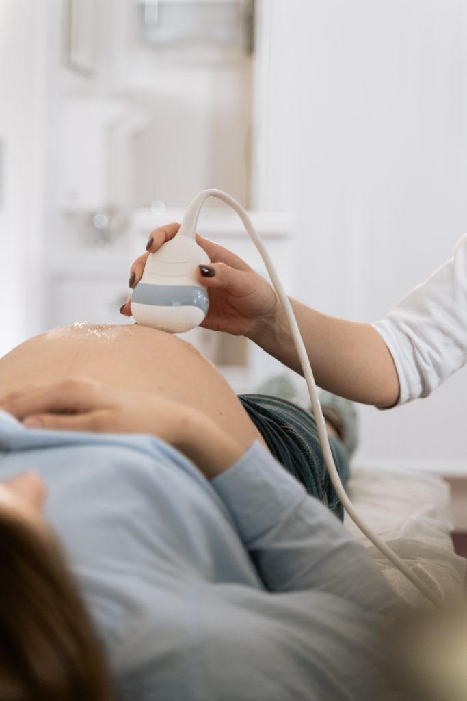
The first ultrasound, or sonogram, will take place when the mother is around six to eight weeks pregnant. At this stage, the baby is still very small and the female’s uterus and fallopian tubes are located closer to the birth canal than to the abdomen. Therefore, the OB/GYN will do this test transvaginally to get a clearer picture. A wand-like transducer probe that emits high-frequency sound waves will be used to create a snapshot of the fetus. The sound waves bounce off of the fetus and send signals back to a machine that produces the reflections into a black and white image. This ultrasound is important because the OB/GYN will listen to the baby’s heart rate and estimate their age by measuring his or her length from crown to rump. This test will allow the doctor to establish a more accurate due date and to better track pregnancy milestones.3
Pregnant females are offered the opportunity to have a nuchal translucency (NT) test performed between eleven and thirteen weeks. The NT involves another ultrasound and evaluates the risk of having a baby with Down syndrome or certain heart defects. The two-part exam consists of a blood test that measures levels of certain proteins and hormones in the mother’s body and includes an ultrasound that shows the thickness of the baby’s neck. Heightened thickness suggests increased risk for birth defects like Down syndrome. The ultrasound will most likely be done on the mother’s abdomen.3
2nd Trimester (Weeks 13 to 26)
During the 2nd trimester, the mother experiences symptoms from the 1st trimester in addition to some new changes. The fetus grows rapidly during this time and his or her sex becomes apparent. Around weeks eighteen and twenty, the mother will have an ultrasound called an anatomy scan.
2nd Trimester: Pregnant Female
The 2nd trimester is a little bit more difficult for the mother than the first. Although a lot of the physical symptoms present during the 1st trimester—breast tenderness, morning sickness, and cramping—have started to decline during the 2nd trimester, there are a few new physical changes that the mother will experience.1 She may start to notice that her belly is growing and that she is starting to look more like a pregnant female. She will start to feel the baby moving, which is called “quickening,” and may even begin to feel some Braxton-Hicks contractions.1 Braxton-Hicks contractions are relatively painless contractions experienced throughout pregnancy that are not associated with going into labor. A female usually experiences more Braxton-Hicks contractions as her pregnancy progresses. The mother will also notice that she experiences the urge to urinate more often and her heart is beating more rapidly during mild activity. She may also begin to have heartburn. These symptoms are all very common and normal indications of pregnancy, so there is typically little cause for conern. The mother will probably still have mood swings during this period, but they are not as severe as in the 1st and 3rd trimesters.1
2nd Trimester: Baby
- Week 13: Thirteen weeks into the pregnancy, the baby’s intestines have receded into his or her abdomen from the umbilical cord where they have been growing. The baby has begun to create urine and discharge it into the amniotic fluid. Also at thirteen weeks, tissue that will develop into bone is growing around the baby’s head and inside his or her arms and legs.2
- Week 14: After fourteen weeks, the baby’s arms have almost grown to the length that he or she will be at birth and his or her neck has become defined. Red blood cells are now forming inside the baby’s spleen. The fetus’s sex will be detectable this week or in the next few weeks. For females, ovarian follicles begin developing. For males, the prostate develops. At this time, the baby may be 3.5 inches long from crown to rump and weight about 1.5 ounces.2
- Week 15: The baby grows rapidly fifteen weeks into the pregnancy. The baby’s skeleton is developing bones, which can be seen on an ultrasound in a couple weeks. The scalp hair pattern is developing as well.2
- Week 16: After sixteen weeks of pregnancy, the baby’s eyes now face forward and can move slowly. The ears are close to being in their final position and the fetus may be capable of making sucking motions with his or her mouth. The baby’s movements are starting to be coordinated. By now, the baby may be more than 4.5 inches long from crown to rump.2
- Week 17: At seventeen weeks, the toenails begin to develop. Fat stores begin to develop under the baby’s skin. These fat stores will provide energy and help warm the baby after birth.2
- Week 18: The fetus may be able to hear at eighteen weeks into pregnancy. At this time, the baby may be 5.5 inches long from crown to rump and weigh around 7 ounces.
- Week 19: After nineteen weeks, the baby is covered with vernix caseosa, which is a greasy, cheese-like coating. The vernix caseosa protects the baby’s sensitive skin from getting scrapes, chapping, and being damaged by exposure to amniotic fluid. For females, their uterus and vagina begin to form at this time.2
- Week 21: Twenty-one weeks into the pregnancy, the baby begins to gain more weight. At this time, he or she is able to swallow and becomes more active.2
- Week 23: After twenty-three weeks, the baby begins to have rapid eye movements. The tongue soon develops taste buds. Fingerprints begin to form. For females, the uterus and ovaries are in place and a lifetime worth of eggs has been formed. For males, the testes are descending into the abdomen. Some babies born prematurely during this week of pregnancy may be able to survive with intense medical care.2
- Week 24: After twenty-four weeks, the baby is regularly sleeping and waking. The fetus is about 8 inches long from crown to rump and weighs more than 1 and ⅓ pounds.2
- Week 25: At week twenty-five, the baby has begun to develop the startle reflex. The startle reflex is when the fetus reacts to a sudden loss of support because it feels as though he or she is falling. Also, the baby may begin to respond to familiar sounds through movement.2
- Week 26: The baby has grown fingernails twenty-six weeks into the pregnancy. The baby’s lungs have started to produce surfactant: a substance that allows the lungs’ air sacs to inflate and keeps them from collapsing. At this time, the baby may be 9 inches long from crown to rump and weigh almost 2 pounds.2 The baby’s lungs and nervous system are maturing nicely by the end of the 2nd trimester.2
2nd Trimester: Checkups
Typically between weeks eighteen and twenty of pregnancy, expecting mothers have a detailed ultrasound called an anatomy scan. It generally lasts about 20 to 45 minutes, but takes longer if the mother is carrying multiple children. This test is used to assess the baby’s growth and to ensure that his or her organs are developing correctly. Doctors or technicians can even be asked to point out particular organs to see! This is the most intensive checkup that the fetus will have before he or she is born. The doctor will check the baby’s heart rate and look for abnormalities in their brain, heart, kidneys, and liver. The doctor will also count fingers and toes, check for birth defects, check the placenta, and measure levels of amniotic fluid. An experienced doctor will be able to determine the sex of the baby 95% of the time.3 This ultrasound will usually provide a better picture of the baby. Sometimes, the mother can even get a 3D view of her baby!
Additionally, between fourteen and twenty weeks, the female can have an amniocentesis, which will check for Down syndrome. The mother should consider an amniocentesis if she is a female who is 35 years and older or if either the mother or father has a family history of certain birth defects. This procedure consists of a needle being inserted through the mother’s belly and into the uterus in order to take a sample of amniotic fluid. There is a small chance of miscarriage from an amniocentesis (0.5%).3
3rd Trimester (Weeks 27 to 40)
Weeks twenty-seven to forty are usually difficult for the mother. The fetus is also uncomfortable as he or she has become cramped inside the mother. During these weeks, the baby starts to prepare for birth.
3rd Trimester: Pregnant Female
The 3rd trimester is usually the hardest of the three trimesters for the female. Most mothers begin to feel very uncomfortable because their stomach is growing rapidly. The mother continues to have the Braxton-Hicks contractions, and most females start to get continuous backaches. In the 3rd trimester, females may have swollen feet caused by fluid retention in the lower half of the body.1 The fact that they are so uncomfortable and have such painful back problems makes it hard for many mothers to sleep during this period.1 The lack of sleep adds to their exhaustion and mood swings. The heartburn usually continues during the 3rd trimester, so females should remain conscious of their diet during these three months. Also, most females will start to experience some droplets of fluid secreting from their nipples. The fluid is normal when a mother is close to the end of her pregnancy and her body begins preparing to breast feed.1
3rd Trimester: Baby
Week 32: After thirty-two weeks of pregnancy, the baby’s central nervous system has matured and begins to regulate body temperature. The baby’s lungs have not fully formed yet, but he or she can now practice breathing. At this time, the fetus could be 11 inches long from crown to rump and weigh 3.75 pounds.
Week 33: At thirty-three weeks of pregnancy, the baby’s pupils can constrict and dilate and they are able to detect light entering their eyes.2
Week 36: The 3rd trimester is also one of the more uncomfortable trimesters for the baby. At thirty-six weeks of pregnancy, the space in the uterus becomes cramped as the baby grows and the mother will notice more sharp movements. The baby will also begin to turn upside down in the uterus.2
Week 37: Thirty-seven weeks into pregnancy, the baby is considered early term. Additionally, all of the baby’s organs function on their own. His or her head may begin to descend into the mother’s pelvis to prepare for birth.2
Week 39: At thirty-nine weeks of pregnancy, the testes of males descend into the scrotum. The placenta continues to give the baby antibodies to combat infection after birth. Breastfeeding babies will provide additional antibodies. The baby’s eyes will start to function better, and he or she will be able to sense changes in light. All the parts of the baby, including the hair and fingernails, continue to grow and develop. By the end of the 3rd trimester, the baby will be ready for life outside his or her mother’s uterus.2
Week 40: After forty weeks of pregnancy, the baby’s due date arrives. The baby may be about 18 to 20 inches long and weigh 6.5 pounds or more. Remember that healthy babies come in a variety of different sizes.2
3rd Trimester: Checkups
For most mothers, their last ultrasound is the anatomy scan that takes place during the twentieth week (2nd trimester) of pregnancy. If the mother’s due date has passed, a doctor may want to closely observe the fetus’ progress with heart rate monitoring and ultrasounds. Other reasons for 3rd trimester ultrasounds include concerns about the placenta, the baby’s growth, low levels of amniotic fluid, bleeding, and pre-term contractions. Additionally, the female will receive a follow-up scan if her cervix was covered by the placenta at her twentieth week scan.3
If the mother has gestational diabetes, she may be scanned with a Doppler ultrasound in the last few weeks of her pregnancy. This type of ultrasound bounces high-frequency sound waves off of circulating red blood cells in order to measure blood pressure and blood flow. The Doppler ultrasound will help show if the baby is receiving enough blood.3
Pregnancy Over the Trimesters

The baby and the mother’s body will change from week to week. All of the symptoms and changes mentioned above are natural and expected for a pregnant female and her fetus. If a female is pregnant and experiences abnormal abdominal pains or cramps, spotting, or extremely intensified versions of the symptoms described above, she should consult a doctor as soon as possible. A pregnant female should also make sure to get regular examinations during the entirety of her pregnancy.
References
1. “Stages of Pregnancy.” Parenting. Meredith Corporation. Web. 18 Jan. 2017.
2. “Pregnancy Week by Week.” Mayo Clinic. Web. 18 Jan. 2017.
3. O’Brien, T. “Ultrasound: A Trimester-by-Trimester Guide.” Parents. Web. 18 Jan. 2017.
Last Updated: 14 February 2017.