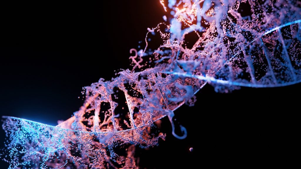
In humans, biological sex is determined by a specific set of chromosomes. Chromosomes are long, threadlike structures of DNA that encode for different traits in an organism. Humans have a total of 46 chromosomes. These include 22 pairs of autosomes, which are the chromosomes that are the same in males and females, and one pair of sex chromosomes, or allosomes, which are different in males and females. Females have two X chromosomes (XX), whereas males have an X and a Y chromosome (XY). An adult female will ovulate around the fourteenth day of her menstrual cycle. Each time a female ovulates, she releases an egg, which contains one X chromosome along with 22 autosomes. An adult male will produce sperm with either an X chromosome or a Y chromosome. Semen, which contains sperm, is released during ejaculation. When an egg and a sperm fuse during reproduction, the chromosome that the sperm carries determines the sex of the child. Sometimes, an individual may receive an abnormal amount of chromosomes, such as in the case of Turner’s Syndrome where females have only one functional X chromosome (XO) and the other sex chromosome is either missing or structurally altered. You can learn more about people who are born with reproductive organs and anatomy that does not fit the typical definitions of female or male here.

Human Sex Differentiation
During the process of sex differentiation, a fetus gains characteristics of either a male or a female. Sex differentiation is initiated and controlled by gonadal steroid hormones. These hormones perform organizing functions to permanently differentiate sex organs during development. This process starts before the developing child is even old enough to be considered a fetus, and is instead still an embryo. A developing human is not considered a fetus until the 9th week of development in the uterus, whereas sex differentiation begins during the 6th week of pregnancy.
By the sixth week of development, all embryos have both Wolffian ducts and Müllerian ducts. The Wolffian ducts are embryonic structures that can form the male internal reproductive system. The Müllerian ducts are embryonic precursor structures to the female internal reproductive system. In this stage, the internal reproductive organ precursors are bipotential, meaning they have the potential to develop into both male and female sex organs given the proper chemical instructions. The way they develop is influenced by hormones, and each fetus will only have one of pair of these ducts by the end of differentiation.2
In Males
For males, the differentiation process is started by the sex determining region Y gene, also known as the SRY-gene on the Y chromosome. This gene generates the necessary biochemistry inside of a male fetus for him to develop male sex organs. The embryonic gonads secrete a protein called the anti-Müllerian hormone, which causes the the Müllerian ducts to degenerate. It also causes the Wolffian ducts to develop into the vas deferens and the seminal vesicles. The undifferentiated gonads develop into testes, and other structures such as the prostate gland and the scrotum develop. This illustration shows the location of the SRY-gene on the Y chromosome.6
In Females
Females have two X chromosomes, so their sexual differentiation is not signaled by the SRY-gene. Instead, the absence of these cues signal their sex organs to develop. The Wolffian ducts degenerate and the Müllerian ducts persist to form the fallopian tubes, uterus, uterine cervix, and the superior portion of the vagina. The undifferentiated gonads develop into ovaries, and other structures such as the labia and vagina develop.6

Development of the External Sex Organs
The external genitalia begin to differentiate during the ninth week of embryonic development and are fully differentiated by the twelfth week. The same initial tissues and structures differentiate to make up the different structures in male and female external genitalia. The illustrations below demonstrate how undifferentiated embryonic external genitalia become different structures in males and females. During embryonic development, the genital tubercle elongates to form a primordial phallus that will either become the clitoris in females or the glans penis in males. The embryonic urethral fold becomes the labia minora in females and the spongy urethra and the ventral aspect of the penis in males. Also, the embryonic labioscrotal swelling becomes the labia majora in females and the scrotum in males.4
Medical Conditions
In some cases, fetuses do not develop as described above. There can be many causes for these differences in development, such as a different genotype, environmental factors, hormone imbalances, and others. The following are three examples of hormonal conditions that change the way that the reproductive organs develop in males or females.
Androgen Insensitivity Syndrome
Androgen insensitivity syndrome (AIS) can occur in both males and females. However, this syndrome has a much more significant impact on males than it does on females. People with this syndrome have tissues that lack sensitivity to androgens, meaning they do not respond as they should to the hormones that cause masculinization. The severity of AIS varies, so development varies widely from person to person. Because the testes secrete anti-Müllerian hormone and the Wolffian ducts do not receive the proper signals to develop, both duct systems degenerate. In the most extreme cases, males will develop external genitalia that resembles the genitalia of a female. They do not have a uterus and therefore cannot menstruate or conceive a child. These genetic males are typically raised as females and have a female gender identity.3
Congenital Adrenal Hyperplasia
Congenital adrenal hyperplasia (CAH) is a condition in which females have overactive adrenal glands. Located above the kidneys, adrenal glands are endocrine glands that produce a variety of hormones. CAH causes excess production of cortisol, a hormone that is structurally and functionally similar to testosterone. The recessive genes responsible for CAH can also be found in males, but they do not have a significant impact on male sex differentiation. Females will develop masculinized genitalia to varying degrees. Some females will have an enlarged clitoris similar to penis formation. Others may have partially fused labia, similar to a scrotum. If it is found early and treated with hormonal therapy, females can develop typical female external genitalia.5
5 α–Reductase Deficiency
Males with 5 α–reductase deficiency (5-ARD) do not undergo the same prenatal sexual differentiation as other males. The enzyme 5 α–reductase helps to regulate proportions of male sex hormones in the body before birth and during puberty. When males have a deficiency of 5-ARD, they will have typical development of the testes, but their external genitalia will resemble that of a female until puberty. During puberty, males will have levels of testosterone that are elevated enough to experience the same changes as males without this condition. After puberty, their genitalia do not look fully masculinized, though many are reported to live perfectly normal male lives.1

References
- Azzouni, F et. al, “The 5 Alpha-Reductase Isozyme Family: A Review of Basic Biology and Their Role in Human Diseases”. Advances in Urology, 2012, 1-18.
- Hannema S, E, Hughes I, A. “Regulation of Wolffian Duct Development”. Hormone Research in Paediatrics, 67, 3, 2007, 142-151.
- Hughes, A et al. “Androgen insensitivity syndrome”. The Lancet , 380, 9851, 2012, 1419 – 1428.
- Moore, K et al. The Developing Human: Clinically Oriented Embryology. Philadelphia, PA: Saunders/Elsevier, 2008. Print.
- Speiser, P, White, P. “Congenital adrenal hyperplasia”. New England Journal of Medicine, 349, 2003, 776-788.
- “SRY gene”. US National Library of Medicine, 2015, 1-5.
Last Updated: 3 November 2016.
