Note: This article was previously titled ‘An Overview of the Female Reproductive System.’ We have changed the language from ‘female’ to ‘people with vaginas’ in the title and throughout the article to be more inclusive, as not all people who identify as women or female have vaginas and not all people with vaginas identify as women or female. Sex and gender are two discrete categories and do not always overlap. If you would like to learn more about gender identity, please visit our article Sexual Orientation and Gender Identity.
The reproductive, or genital, systems of people with vaginas are made up of external and internal sex organs. These sex organs, each of which have their own complex functions, work together to provide the body sexual pleasure and reproductive abilities. This article offers an overview of the sex organs and their functions, beginning with external anatomy and moving on to the internal anatomical structures.
Table of Contents
I. The Vulva
The vulva refers to the entirety of a person with a vagina’s external genitalia. The vulva consists of the following structures: the (a)Mons Pubis, (b)Clitoral Hood, (c)Glans Clitoris, (d)Urethral Opening, (e)Vestibule, (f)Labia Majora, (g)Labia Minora, and (h)Vaginal Opening.
a. Mons Pubis – The mons pubis, often referred to as solely ‘the mons,’ is the upper and frontmost component of the vulva.1 It is a pad of fatty tissue that is covered by skin and pubic hair (which often first appears during puberty) and serves both sexual and physiological functions. It may be sexually arousing for a partner as it is the most visual part of the vulva. It can also serve as a cushion during sexual activity. The pubic hair serves to vaporize odors produced by sweat glands in this area.
b. Clitoral Hood – One of the many components of the clitoris, the clitoral hood is a loose fold of skin that fully or partially covers the external portion of the clitoris, the (c)glans clitoris. It is formed by the joining of the frontmost portion of the left and right (g)labia minora.
c. Glans Clitoris – The glans, or the external portion of the clitoris, is a small but incredibly sensitive knob of tissue found at the front of the (e)vestibule. It is an erogenous zone that contains more nerve endings than any other body part. Like most other parts of the vulva, the glans clitoris can also vary in size, shape, and sensitivity3.
d. Urethral Opening2 – This is the external opening of the urethra, a tube in which the urine leaves the body from the bladder.
e. Vestibule – This is the smooth surface located between the left and right (g)labia minora which begins just below the (c)clitoris and ends at the bottom of the labia minora.1 It contains the (d)urethral and (h)vaginal openings.
f. Labia Majora – Also known as the ‘outer labia’ or ‘outer lips,’ the labia majora are the pair of fleshy skin folds that extend from the (a)mons. The outer portion of this area nearest the thighs is often covered in pubic hair and can appear darker than other areas of the vulva. This area is erotically sensitive, especially the inner, hairless portion.
g. Labia Minora – The ‘inner labia’ or ‘labia minora’ are a pair of thin folds of hairless skin that is found in between the (f)labia majora.1 This portion of the vulva is highly variable in size, shape, and color. The labia minora are often incredibly erotically sensitive, as they have a rich supply of blood vessels and nerve endings. During arousal, increased blood supply causes this area to swell and darken, a process known as vasocongestion.
h. Vaginal Opening2 – This is the external opening to the vagina. It is the muscular outermost portion of the internal reproductive tract that forms the birth canal and is the receptor for the penis during coitus.
II. The Clitoris
The clitoris refers to a group of structures, both internal and external. It is made up of the: (b)Clitoral Hood, (c)Glans Clitoris, (i)Corpus Cavernosum, (j)Bulbs of Vestibule (or vestibular bulbs), and (k)crus.
The structures, locations, and functions of the clitoral hood and glans clitoris are outlined above.
i. Corpus Cavernosum – This term is the singular form of the corpora cavernosa, a set of two internal elongated erectile structures within the clitoral shaft that lie side by side and become erect with sexual arousal.
j. Bulb of Vestibule – This is one of two curved masses of erectile tissue that surround the (e)vestibule and are found internally, underlying the inner labia.1 Like the (i)corpora cavernosa, the vestibular bulbs also become engorged with blood during sexual arousal.
k. Crus – This is the term for the singular form of “crura”, the portion of the (i)corpora cavernosa in which the two sides diverge from one another, forming a left and a right side. This gives the internal clitoris a wishbone structure.1
III. The Vagina and Related Structures
The vagina is a stretchable muscular cavity lined with mucous membranes that extends from the vulva to the cervix. The vagina includes the following structures: (h) Vaginal Opening and Vaginal Canal, (l)Skene’s Glands, and (m)Bartholin’s Glands.
l. Skene’s Glands – The paraurethral glands, or the Skene’s glands, are located on the vaginal walls next to the urethra. It is theorized that they are potentially also part of the G-spot4. The paraurethral glands also include the clitoris and swell up with blood during sexual arousal. Ejaculation may occur in the paraurethral glands. The paraurethral glands are incredibly sensitive and, when stimulated properly, can produce an orgasm in some individuals.
m. Bartholin’s Gland2 – The Bartholin’s Glands are two glands located towards the back end of the vestibule that secrete mucus which assists with lubricating the vaginal opening.
IV. The Uterus and Related Structures
The uterus is a portion of the internal anatomy that plays a large role in menstruation, fertilization, and pregnancy. The following structures can be found in and around the uterus and are largely responsible for the aforementioned processes: (n)Fallopian Tubes, (o)Uterine Cavity, (p)Eggs, (q)Fimbria, (r)Ovaries, (s)Endometrium, (t)Cervix, and (h)Vagina.
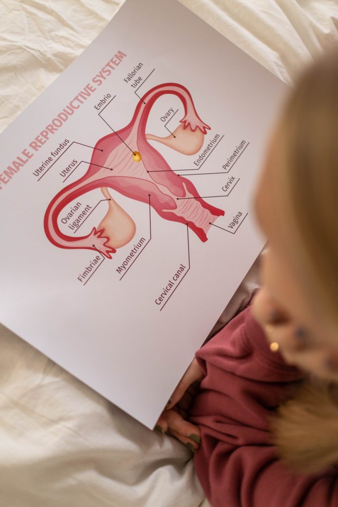
n. Fallopian Tube(s) – Also known as ‘oviducts’ or simply as ‘tubes,’ the fallopian tubes are two tubes, each about 4 inches long, that form a pathway between the uterus and the left and right ovaries. Fertilization of an (p)egg by sperm occurs in this pathway, which is why the process of a person with a uterus undergoing a tubal ligation7 (or having their ‘tubes tied’) is so effective at preventing pregnancy.
o. Uterine Cavity (Uterus) – The uterus, also referred to as the “womb,”, is a hollow organ that is found within the pelvic cavity and beyond the vagina3. It is made up of three layers, the innermost of these being the endometrium, which sheds during menstruation and is where fertilized eggs implant. The middle layer, or the myometrium, is a wall composed primarily of smooth muscle, whose main function is to contract during childbirth. The perimetrium is the tough outer layer, which separates the uterus from the pelvic cavity and protects the fetus. In people who are not pregnant, the uterus is typically the shape and size of a small upside-down pear. However, during pregnancy, it expands to allow for the growth of the fetus.
p. Egg(s) – Eggs, or more formally ‘ova’ are the gametes that are fertilized by sperm during fertilization, resulting in pregnancy. Eggs are stored in the (r)ovaries and released in a cycle known as ‘ovulation6’, which is the most fertile period of the menstrual cycle.
q. Fimbria – The fimbria are the finger-like projections located at the ends of the (n)fallopian tubes, on the ovary side of each oviduct. Their function is to to catch the ovum, or (p)egg, after is is released from the ovaries.
r. Ovaries – These are a pair of egg-shaped organs whose function is to release a mature ovum (egg) during ovulation. They are also responsible for the production and secretion of sex hormones6. They are located on either side of the (o)uterus, near the (q)fimbria3.
s. Endometrium6 – This is the innermost lining of the uterus. It is the portion that is shed during menstruation if fertilization does not occur and the site where fertilized eggs implant if it does3.
t. Cervix – This is the lowermost, narrow portion of the uterus that connects with the vagina.1 It is often able to be felt by inserting fingers into the vaginal canal. It serves to separate the uterus and the vagina, and during pregnancy, holds the fetus in the uterus until delivery, at which point it dilates to assist with childbirth5. The cervix also contains the ‘Os1’ which is the constricted opening that connects the vagina to a short canal which opens into the uterus.
V. Structural Organs
The structural organs hold the entire reproductive system in place. These structures include: (u)Pelvic Floor Muscles and the (v)Pubic Bone.
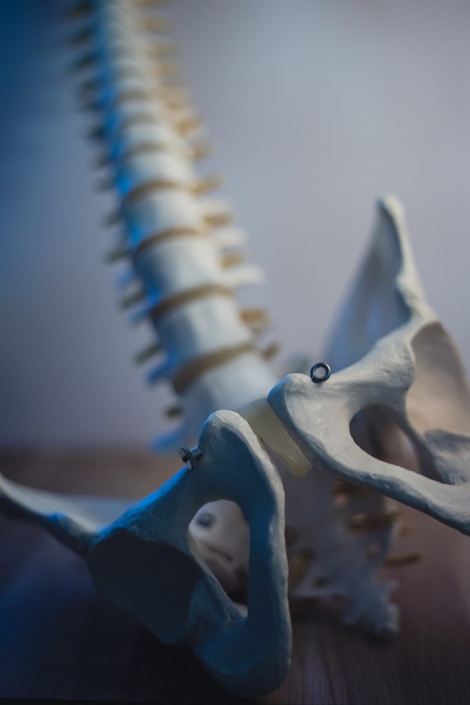
u. Pelvic Floor Muscles – The pelvic floor muscles compose a muscular sling that underlies and supports the organs located in the pelvis. Specifically, the PC muscle, or pubococcygeus muscle, is the muscle of the pelvic floor that spans from the pubic bone to the tailbone.1 Doing Kegel exercises can help maintain a strong PC muscle, which increases sexual stimulation and can also help with urinary and fecal control.
v. Pubic Bone2 – Located under the (a)mons, the main function of the pubic bone, or pubis, is to protect the organs that lie underneath, specifically the intestines, bladder, and internal sex organs. It also helps with the movement and the stabilization of the pelvis.
VI. Related Non-Reproductive Structures
The following structures are located in the same general area, but do not have reproductive functions. The non-reproductive structures include: the (w)Rectum, (x)Bladder, (y)Anus, and (z)Perineum.
w. Rectum – The rectum is the lower part of the large intestine where stool is stored before it passes out of the body via the anus. It is a continuation of the colon and connects to the exterior of the body via the (y)anus.
x. Bladder – The bladder is a hollow organ located behind the pubic bone in which urine is stored prior to being expelled through the (d)urethra. Kegel exercises can help to strengthen the muscles involved in the passage of urine, thus leading to greater bladder control and less unintended leakage.
y. Anus – This is the external opening of the rectum from which feces is discharged. It is also the structure used in “anal sex,” or the manual, oral, or penetrative stimulation of the anus1. Due to a higher frequency of bacteria, special care should be taken when participating in anal sex in order to avoid transmission of sexually transmitted infections (STIs).
z. Perineum – This is the strip located between the (h)vaginal opening and the (y)anus. It is often erotically sensitive, but is also a pathway for anal bacteria to travel to the vagina, which is why people with vaginas are advised to wipe front-to-back.1
Concluding Remarks
The more knowledge one has about their reproductive organs, the less daunting reproductive and sexual issues may appear. Additionally, awareness of a healthy reproductive system allows one to make informed decisions regarding their physical, emotional, and sexual lives.

References
- LeVay, S., Baldwin, J., & Baldwin, J. “Women’s Bodies.” Discovering Human Sexuality (5th ed.). Oxford University Press. 2021.
- “NCI Dictionary of Cancer Terms.” Cancer.gov. National Cancer Institute.
- “Female Genital Anatomy.” Bumc.bu.edu. Boston University School of Medicine.
- Puppo, Vincenzo. “Embryology and Anatomy of the Vulva: The Female Orgasm and Women’s Sexual Health.” Elsevier.com. European Journal of Obstetrics and Gynecology and Reproductive Biology, 13 Aug. 2010.
- Nguyen, John. “Anatomy, Abdomen, and Pelvis, Female External Genitalia.” Ncbi.nlm.nih.gov. Florida Atlantic University, 31 Jul. 2021.
- Campbell, Kenisha. “Menstrual Cycle: An Overview.” Chop.edu. Children’s Hospital of Philadelphia.
- “Tubal Ligation.” Hopkinsmedicine.org. John Hopkins Medicine.
Last Updated: 3 March 2022
